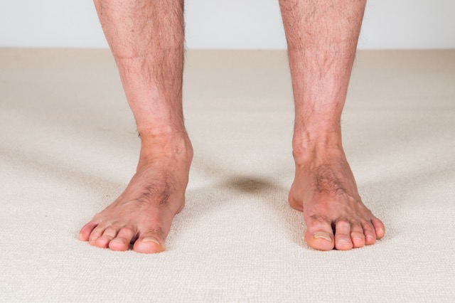
Introduction to overpronation
Pronation and supination are normal healthy movements of the foot that primarily occur at the subtalar joint between the talus and calcaneus (note: it also occurs at the transverse tarsal joint, which itself is composed of two separate joints: the talonavicular joint and the calcaneocuboid joint). These movements occur in one oblique plane around one axis, therefore they are uniaxial; however, because the oblique plane movement occurs across all three cardinal planes, pronation and supination are often described as triplanar motions. The principle cardinal plane component motion of pronation is frontal plane eversion. However, pronation also involves subtalar abduction (effectively lateral rotation) of the foot in the transverse plane and subtalar dorsiflexion of the foot in the sagittal plane.
Note: The term pronation is not synonymous with eversion; rather, the cardinal plane component motion of eversion is the principal component of the oblique plane motion of pronation. Similarly, the term supination is not synonymous with inversion; rather, the cardinal plane component motion of inversion is the principal component of the oblique plane motion of supination.
Foot pronation causes the arch structure of the foot to drop. The arch structure consists of three arches: the medial longitudinal arch on the big toe side, which is the largest of the arches; the lateral longitudinal arch on the little toe side; and the transverse arch across the metatarsal heads. Whenever any one of these arches collapses, as a rule, the entire arch structure collapses.
Because overpronation causes the arch structure to drop, it is known in lay terms as flat foot. There are two types of overpronation/flat foot: rigid flat foot and supple flat foot. With supple flat foot, which is the more common of the two, the client’s/patient’s arch is perfectly healthy when they are not weight bearing, but upon weight bearing, the foot pronates excessively and their arch collapses. By contrast, a rigid flat foot is always flat/overly pronated, regardless of whether the client/patient is weight bearing or not.
Causes
There are many causes of supple flat foot. Given that the arch structure of the foot is determined by soft tissue pulls of musculature and fascial tissue (ligaments and joint capsules), a supple flat foot is caused by either lax ligaments and/or weak musculature that cannot support the arch when the weight of the body passes through the subtalar joint. Muscles that act to support the arch can be divided into the following groups:
- Supinators (invertors) of the foot – These muscles have their bellies located in the leg. They are the tibialis anterior and the extensor hallucis longus in the anterior compartment; and the Tom, Dick and Harry group: tibialis posterior, flexor digitorum longus, and flexor hallucis longus muscles of the posterior deep compartment.
- “Stirrup Muscles” – The stirrup muscles, also located in the leg, are named because they support the arch/underside of the foot like a stirrup. They are tibialis anterior (already mentioned above) and the fibularis longus.
- Intrinsic plantar musculature – Layer I of the intrinsic plantar musculature have attachments into the plantar fascia; by supporting the plantar fascia, they help to support the arch. They are the flexor digitorum brevis, abductor hallucis, and abductor digiti minimi pedis.
- Lateral rotators of the thigh at the hip joint – This group indirectly supports the arch because it acts to prevent the thigh from medially rotating. When the weight bearing foot pronates, because the foot is planted on the ground, the calcaneus of the subtalar joint is not free to fully move, therefore the talus moves as well. This is a closed-chain reverse action of the proximal talus upon the distal calcaneus, and results in medial rotation of the talus. Because the ankle joint does not allow rotation, the tibia medially rotates with the talus; and because the extended knee joint also does not allow rotation, the thigh medially rotates with the leg. Therefore, hip joint lateral rotation musculature can support the arch by acting to brake/prevent medial rotation of the thigh/leg/talus. Hip joint lateral rotation musculature includes the posterior gluteal musculature, the deep lateral rotator group (piriformis, quadratus femoris, superior and inferior gemellus, and obturator internus and externus) and the sartorius.
- It should also be mentioned that hip joint abductor musculature can also be important for maintaining the arch of the foot. If this musculature is weak, the thigh can fall into adduction, this causes a genu valgus force (abduction of the leg at the knee joint), which tends to result in medial rotation of the thigh, and therefore the leg and talus, promoting arch collapse. Abductors of the hip joint are the gluteal muscles, tensor fasciae latae (TFL), and the sartorius.
Another contributor to overpronation is tight pronator (evertor) musculature (fibularis musculature and extensor digitorum longus), which can pull the foot into pronation on that side, making it more difficult for the supinator musculature to support the arch structure.
Most all ligaments that are located on the plantar side of the foot help to support the arch. Most notable are the long and short plantar ligaments, the spring ligament, and the intertransverse metatarsal ligaments. If the ligaments are excessively lax, perhaps due to genetic factors or to forces placed upon them during life, they will not be able to hold the bones in their proper posture, especially during weight bearing, and the arch will collapse.
As stated, the collapsed arch of overpronation essentially occurs because of the inability of the musculature and fascial ligament complex to support the arch, especially when weight bearing. Therefore, any factor that increases downward force through the arch will tend to exacerbate this condition. First among them is being overweight, which increases the weight that is borne through the arch. Carrying heavy loads/objects acts in a similar manner because the weight of whatever is being carried must ultimately pass through the arches.
Another factor is a turned out posture of the foot. This usually occurs because of excessively tight baseline tone of deep lateral rotation musculature of the thigh at the hip joint (e.g., piriformis). When walking with a turned out posture, the person’s weight passes more directly over the arch, increasing the likelihood that it will collapse. Ironically, the baseline tone of the lateral rotation musculature of the hip joint might be tight enough to cause the unhealthy turned out posture of the foot, but not strong enough to prevent the foot from overly pronating as a result of this altered posture. It is important for the lateral rotation musculature to have a healthy and loose baseline tone, but to be strong enough to contract to prevent overpronation when needed during the gait cycle.
Proper footwear can be another factor. If a person does overly pronate, then wearing shoes that have little or no arch support can allow the excessive pronation to occur. Wearing high-heeled shoes can also exacerbate this problem because they shift body weight to be borne more anteriorly in the foot, increasing force through the transverse arch, causing it to collapse. This will result in weakness of the entire arch structure of the foot, including the medial longitudinal arch and over pronation.
Finally, the longer that a client/patient has had an overly pronated foot, the more likely it is that fascial adhesions accumulate that exacerbate the condition by holding the foot in a posture of excessive pronation. This is especially true for rigid flat foot, but might become a factor that causes a supple flat foot to gradually become a rigid flat foot.


