Signs and symptoms:
The most common symptom of Achilles’ tendinitis is pain at the Achilles tendon. Pain is usually not present at rest, but will be evident upon palpation. Pain will also be provoked if the triceps surae musculature contracts to plantarflex the foot or is stretched into dorsiflexion because both of these circumstances will result in a pulling/tensile force being placed on the tendon. Inflammation and crepitus also often occur with Achilles tendinitis. Because Achilles tendinitis is caused by overuse of the triceps surae musculature, myofascial trigger points and/or global tightness can usually be found in the gastrocnemius and soleus. Plantarflexion is used whenever we walk; for this reason, it is difficult to rest the triceps surae musculature, so Achilles tendinitis can be a very chronic condition.
Associated disorders of the Achilles tendon
Achilles bursitis and paratendinitis also exhibit inflammation and tenderness, especially with use of the Achilles tendon; whereas Achilles tendinosus does not involve inflammation. Signs and symptoms of a ruptured Achilles tendon are much more dramatic. The client/patient will not be able to plantarflex the foot with any strength because of the loss of use of the gastrocnemius and soleus. Palpation will reveal a gap where the tendon is supposed to be. Because of the inherent tension with the gastrocnemius and soleus, when the tendon is ruptured, the muscle bellies will be pulled up toward their proximal attachments and they can be palpated and most likely visually seen to be “balled up” near the knee joint. The client/patient will also often recall a loud popping noise and/or a sudden severe sharp pain at the back of the foot.
Assessment/Diagnosis:
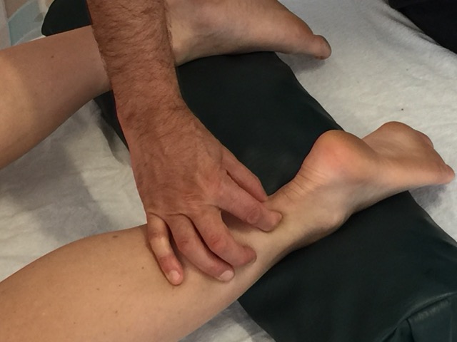
Assessment/diagnosis of Achilles tendinitis is made by correlating history with physical examination findings. The client/patient will usually relate that they had an increase in physical activity that involves plantarflexion of the foot prior to the onset of symptoms. They will also report pain at the tendon when plantarflexing the foot against resistance as well as when the foot is actively or passively dorsiflexed at the ankle joint. Dorsiflexion range of motion will most likely be decreased. The Achilles tendon pinch test is performed by pinching the medial and lateral sides of the Achilles tendon, superior to the location of the bursae. The presence of pain usually confirms tendinitis. Swelling will be palpably and visibly present. The gastrocnemius and soleus should also be palpated for myofascial trigger points and global tightness. Because plantarflexion against resistance and active and passive dorsiflexion are painful, the client/patient will usually exhibit an antalgic gait.
Associated disorders of the Achilles tendon
The Achilles pinch test will also show positive for Achilles paratendinitis. If performed more distally at the level of the Achilles bursae, the positive pinch test may also be indicative of bursitis as well as tendinitis.
A rupture of the Achilles tendon is assessed by the history of a loud popping noise and/or a sudden severe pain when plantarflexing, as well as a gap where the tendon should be located near the calcaneus, bunching up of the gastrocnemius and soleus bellies proximally, and an inability to plantarflex against resistance. The triceps surae squeeze test, also known as the Thompson test can be used to assess Achilles rupture. With the client/patient prone and the foot hanging off the table, the therapist squeezes on each side of the triceps surae group near the midpoint of the leg. This action should result in some passive plantarflexion. The absence of plantarflexion is indicative of Achilles tendon rupture.
Differential assessment:
Differential assessment of Achilles tendinitis involves the consideration of Achilles tendinosus, bursitis, paratendinitis, and rupture. Other conditions that should be kept in mind are global tightness of or myofascial trigger points within the gastrocnemius and/or soleus; these conditions can also result in pain with plantarflexion against resistance and pain and decreased dorsiflexion of the foot. And whenever there is pain in the posterior (lower) leg, deep vein thrombosis should be ruled out.


